
A close look into the mind of a sheep
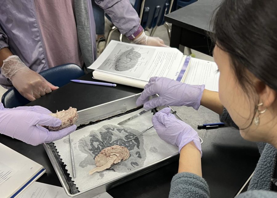
Sophia Avellaneda and Haven Major work together to slice into a sheep brain in Andrew Bedinger’s 2B Anatomy class on Feb. 2, 2023
March 6, 2023
30 minutes.
That’s all it took for five anatomy classes to complete one of their five yearly dissections.
Although the dissection itself is quick, it left a lasting impact on many students.
“As someone who is very passionate about going into the medical field in the future, the dissection really helped me get the experience necessary for my career. It truly helped me visualize the anatomy of the brain in a different way,” said junior Iana Niknezhad.
Students begin by dissecting the outside of the sheep brain’s protective coverings called the meninges. Next, they observe the cerebrum, brainstem and cerebellum and try to locate where the 12 cranial nerves are attached to the brain. On the inside, they look for cavities of white and gray matter which are normally filled with cerebrospinal fluid.
“The main benefit of the dissection was the hands-on experience. Being able to dissect the brain and see the individual parts was much more helpful than just photos and diagrams,” said junior Brandon Cheng.
Students found that the most challenging part of the dissection was getting the innermost layer of meanings, also known as pia mater off of the brain. Although, through collaboration and with the help of dissecting scissors, a dissecting needle, and blunt probe, they were able to carefully remove the pia mater.
“While the students may be a long way from performing surgery, it allows them to practice a skill and go about the learning in a manner that is very different from anything else they do in school,” said science teacher Andy Bedinger. “Dissections are such a unique experience that students can hardly contain their excitement.”